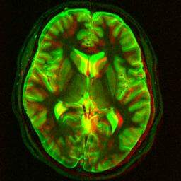
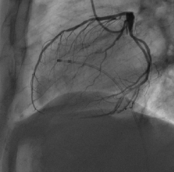
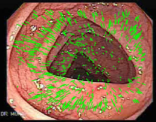
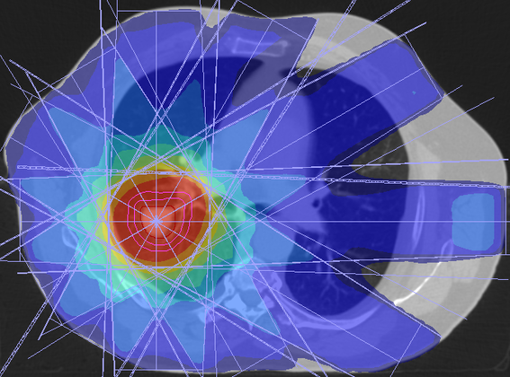
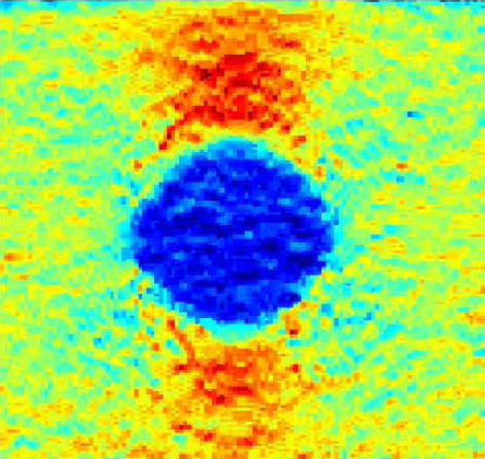
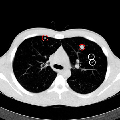
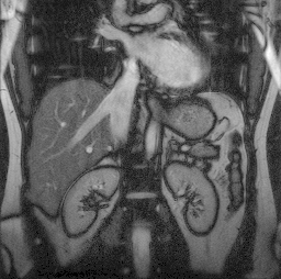
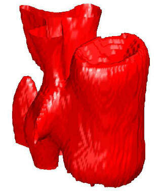
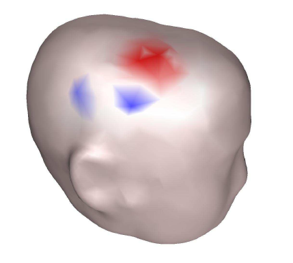
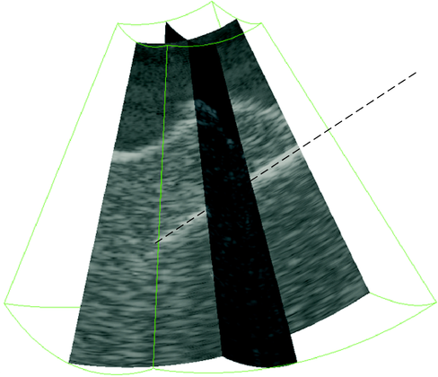
The amount of image data produced in medicine is growing very rapidly. Many previously analogue modalities (e.g. X-ray) now provide digital data, modalities providing 3D data (e.g. MRI or CT) are already routinely used in everyday clinical practice and their resolution is increasing year by year, thus increasing the size of the data produced.
Today, the speed with which a patient learns the result of an examination is no longer limited by technology, but by the availability of radiologists. Many hospitals around the world routinely send their data to China and India. The group is developing tools to make doctors' jobs easier and faster, for example by highlighting changes since the last scan or highlighting areas of potential interest. In the future, the computer will then be able to make a diagnosis completely independently, and in some fields it already has better results than the average doctor
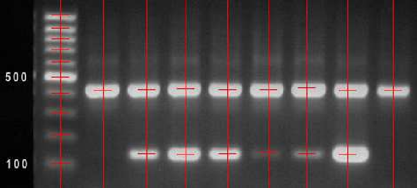
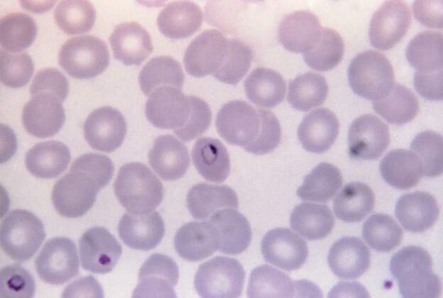
Even in biology, the volume of data generated is growing at a dizzying pace - 3D microscopy is becoming more common, resolution is increasing, and robotic workstations can automatically prepare and image slides. However, it is not humanly possible, for example, to look at all the cells on a single slide and determine whether each one contains a parasite or a genetic anomaly.
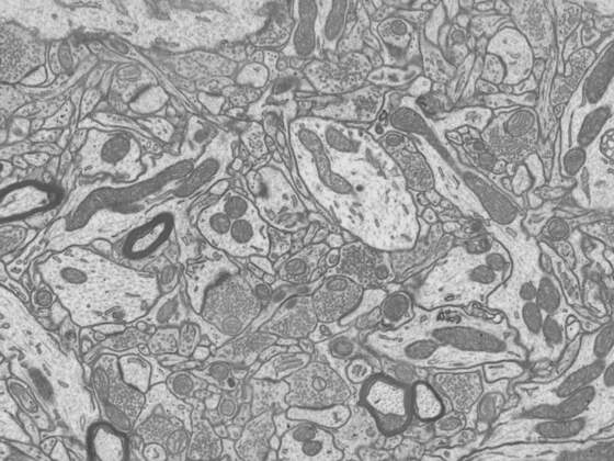
It takes a person months to draw nerve fibres in a piece of tissue smaller than a millimetre, so there is also great potential for computer algorithms that could make the evaluation of scanned data much faster. The advantage is that, unlike medical applications, absolute accuracy is often not required here.


